What are fibroids?
Fibroids are muscular tumors that grow in the wall of the uterus (womb). Another medical term for fibroids is "leiomyoma" (leye-oh-meye-OH- muh) or just "myoma". Fibroids are almost always benign (not cancerous). Fibroids can grow as a single tumor, or there can be many of them in the uterus. They can be as small as an apple seed or as big as a grapefruit. In unusual cases they can become very large.
There are factors that can increase a woman's risk of developing fibroids.
Age. Fibroids become more common as women age, especially during the 30s and 40s through menopause. After menopause, fibroids usually shrink.
Family history. Having a family member with fibroids increases your risk. If a woman's mother had fibroids, her risk of having them is about three times higher than average.
Ethnic origin. African-American women are more likely to develop fibroids than white women.
Obesity. Women who are overweight are at higher risk for fibroids. For very heavy women, the risk is two to three times greater than average.
Eating habits. Eating a lot of red meat (e.g., beef) and ham is linked with a higher risk of fibroids.
Eating plenty of green vegetables seems to protect women from developing fibroids.
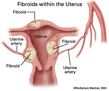
Most fibroids grow in the wall of the uterus. Doctors put them into three groups based on where they grow:
Submucosal (sub-myoo-KOH-zuhl) fibroids grow into the uterine cavity.
Intramural (ihn-truh-MYOOR-uhl) fibroids grow within the wall of the uterus.
Subserosal (sub-suh-ROH-zuhl) fibroids grow on the outside of the uterus.
Some fibroids grow on stalks that grow out from the surface of the uterus or into the cavity of the uterus. They might look like mushrooms. These are called pedunculated (pih-DUHN-kyoo-lay-ted)
fibroids.
Most fibroids do not cause any symptoms, but some women with fibroids can have: Heavy bleeding (which can be heavy enough to
cause anemia ) or painful periods
Feeling of fullness in the pelvic area (lower stomach area)
Enlargement of the lower abdomen
Frequent urination
Pain during sex
Lower back pain
Complications during pregnancy and labor, including a six-time greater risk of cesarean section
Reproductive problems, such as infertility , which is very rare.
No one knows for sure what causes fibroids. Researchers think that more than one factor could play a role. These factors could be:
Hormonal (affected by estrogen and progesterone levels)
Genetic (runs in families)
Because no one knows for sure what causes fibroids, we also don't know what causes them to grow or shrink. We do know that they are under
hormonal control — both estrogen and
progesterone. They grow rapidly during
pregnancy, when hormone levels are high. They shrink when anti-hormone medication is used.
They also stop growing or shrink once a woman reaches menopause.
Women who have fibroids are more likely to have problems during pregnancy and delivery. This doesn't mean there will be problems. Most
women with fibroids have normal pregnancies. The most common problems seen in women with fibroids are:
Cesarean section. The risk of needing a c-section is six times greater for women with fibroids.
Baby is breech. The baby is not positioned well for vaginal delivery.
Labor fails to progress.
Placental abruption. The placenta breaks away from the wall of the uterus before delivery. When this happens, the fetus does not get enough oxygen.
Preterm delivery.
Talk to your obstetrician if you have fibroids and become pregnant. All obstetricians have experience dealing with fibroids and pregnancy.
Most women who have fibroids and become pregnant do not need to see an OB who deals with high-risk pregnancies.
Your doctor can do imaging tests to confirm that you have fibroids. These are tests that create a "picture" of the inside of your body without surgery. These tests might include:
Ultrasound – Uses sound waves to produce the picture. The ultrasound probe can be placed on the abdomen or it can be placed inside the vagina to make the picture.
Magnetic resonance imaging (MRI) – Uses magnets and radio waves to produce the picture
X-rays – Uses a form of radiation to see into the body and produce the picture
Cat scan (CT) – Takes many X-ray pictures of the body from different angles for a more complete image
Hysterosalpingogram (hiss-tur-oh-sal-PIN-juh- gram) (HSG) or sonohysterogram (soh-noh-HISS-tur-oh-gram) – An HSG involves injecting x-ray dye into the uterus and taking x-ray
pictures. A sonohysterogram involves injecting water into the uterus and making ultrasound pictures.
You might also need surgery to know for sure if you have fibroids. There are two types of surgery to do this:
Laparoscopy (lap-ar-OSS-koh-pee) – The doctor inserts a long, thin scope into a tiny incision made in or near the navel. The scope has a bright
light and a camera. This allows the doctor to view the uterus and other organs on a monitor during the procedure. Pictures also can be made.
Hysteroscopy (hiss-tur-OSS-koh-pee) – The doctor passes a long, thin scope with a light through the vagina and cervix into the uterus. No incision is needed. The doctor can look inside the
uterus for fibroids and other problems, such as polyps. A camera also can be used with the scope.